We see more so you can see better
State‑of‑the‑art diagnostic testing powered by compassion and precision.


State‑of‑the‑art diagnostic testing powered by compassion and precision.
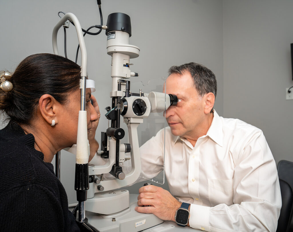
We believe that our cutting-edge technology and high-quality care go hand-in-hand. We offer a variety of advanced technology options to serve the range of your eye care needs in a comfortable and safe environment.
|
Benefits |
Test Name |
What it does? |
■ OHIP |
|---|---|---|---|
|
This test helps predict if you will develop corneal clouding (which can require surgery to fix) before obvious signs show up. |
Endothelial Cell Count |
Measures the quantity and health of the innermost cells in the cornea. |
■ |
|
This test is essential for cataract surgery planning and useful when dense cataracts prevent optical measurements. |
A-Scan |
An A-Scan is an ultrasound test used to measure the length of the eye. These measurements help your surgeon calculate the correct intra-ocular lens (IOL) power needed for cataract surgery. |
■ |
|
The IOL master allows for precise lens selection for cataract surgery; greatly reducing the need for glasses. |
IOL Master |
Measures the exact length of the eye using laser technology that is 10x more accurate than the standard test. |
■ |
|
Corneal curvature can cause vision distortion, measuring it allows more accurate lens choice ahead of cataract surgery. It also helps with uncovering some common inherited diseases. |
Topography |
Measures the shape & curvature of the entire cornea. |
■ |
|
Aberrometry determines which structures in your eye might be scattering lights and causing glare or blurred vision; symptoms that can otherwise be hard to diagnose. |
OPD‑Scan III (Abberometry) |
Tailored treatment plans, fewer side effects like glare or halos, better visual comfort. |
■ |
|
This test helps diagnose and monitor conditions such as keratoconus, assess corneal health, and assist with planning for cataract surgery, laser vision correction, and other eye procedures. |
Pentacam |
Pentacam is a non-invasive imaging test that uses a rotating camera and specialized technology to create a detailed 3D map of the front surface of the eye. It measures the shape, thickness, and curvature of the cornea, as well as the depth of the anterior chamber. |
■ |
|
This helps patients with other vision limiting diseases (like macular degeneration or glaucoma) determine what level of vision they can expect after cataract surgery. |
Potential Acuity Meter (PAM) |
PAM helps determine how much of a patient’s vision loss can be attributed to cataracts. |
■ |
|
OCT helps diagnose and monitor conditions such as glaucoma, macular degeneration, diabetic retinopathy, retinal swelling, and other retinal diseases. The test is quick, painless, and provides highly detailed images that assist in early detection, treatment planning, and monitoring of disease progression. |
OCT (Optical Coherence Tomography) |
OCT is a non-invasive imaging test that uses light waves to create detailed cross-sectional images of the retina and optic nerve. It allows your doctor to see the different layers of the eye and detect subtle changes that may not be visible during a regular eye examination. |
■ |
|
This detailed image helps detect and monitor conditions such as diabetic retinopathy, retinal tears, retinal detachments, macular degeneration, and other retinal diseases. |
Wide-Field Retinal Imaging (OPTOS) |
Optos is a non-invasive imaging test that captures a wide-angle photograph of the retina, allowing your doctor to view up to 200 degrees of the back of the eye in a single image. |
■ |
|
Visual field testing measures your peripheral (side) vision and helps identify areas of vision loss that may not be noticeable during daily activities. It is an important tool for diagnosing and monitoring glaucoma, detecting vision changes caused by retinal or neurological conditions, tracking disease progression over time, and evaluating how well treatment is working. |
Visual Field Analyzer |
Detects and tracks glaucoma progression, protecting peripheral vision before noticeable loss occurs. |
■ |
|
OCT Angiography helps detect and monitor conditions such as macular degeneration, diabetic retinopathy, retinal vascular abnormalities, and other retinal diseases. The test is quick, painless, and provides detailed information about retinal blood flow, helping with early diagnosis, treatment planning, and ongoing monitoring. |
Angiography (OCTA) |
OCT Angiography is a non-invasive imaging test that uses advanced OCT technology to capture detailed images of the blood vessels in the retina and beneath the retina without the need for injections or dye. The scan maps blood flow within the eye, allowing your doctor to evaluate the health of the retinal circulation. |
■ |
|
LipiScan helps diagnose and evaluate meibomian gland dysfunction (MGD), one of the leading causes of dry eye disease. By assessing the health and structure of the glands, the test helps guide treatment recommendations and monitor changes over time, allowing for more targeted and effective dry eye management. |
LipiScan |
LipiScan is a quick, non-invasive imaging test that captures detailed images of the meibomian glands located within the eyelids. These glands produce the oil layer of your tears, which helps prevent tears from evaporating too quickly and keeps the eyes comfortable. |
■ |
See our Patient Uninsured Testing Brochure for more information.
Brampton
7700 Hurontario Street, Unit 605
Brampton, Ontario L6Y 4M3


Mississauga-Oakville
2201 Bristol Circle, Suite 100,
Oakville, Ontario L6H 0J8

©Prism Eye Institute all rights reserved 2026. Designed by Glacial Multimedia.
If you are using a screen reader and are having problems using this website, please call 905-456-3987.